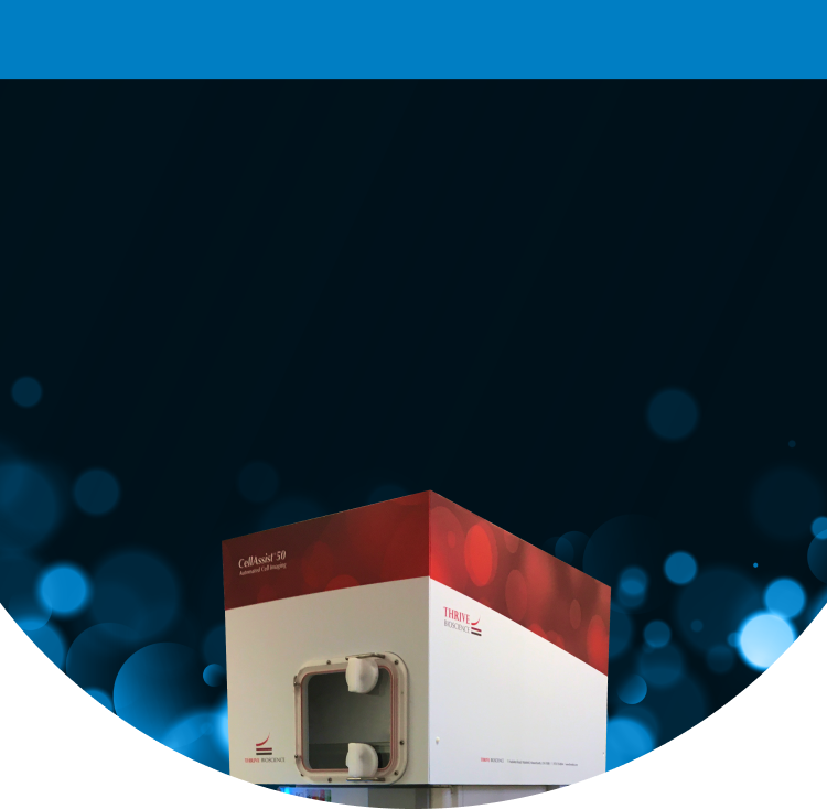
CellAssist 50 System

High-Throughput Live Cell Analysis Platform Designed to Enable Quantitative Cell Biology

Contact Us

Scaling Up Your Cell Culture Experiments with Precision and Efficiency

An automated time series cell imaging system with the same imaging and software that drives the CellAssist Benchtop Imager, paired with an integrated 50-plate CO2 incubator. The CellAssist 50 System is ideal for large-scale experiments, or managing multiple projects and users in parallel. With dynamic scheduling, each of the plates can have separate imaging schedules and imaging modes, allowing for consistently timed around-the-clock experiments.

Maximizing Imaging Throughput with Rich, Reproducible Data

High-Throughput Efficiency

Image up to 50 cell culture plates automatically with up to 400 scans per day, significantly increasing your data collection capacity and saving you valuable time that would otherwise be spent on manual tasks. Superior image registration allows for enhanced tracking and analysis of live cells frequently over time.

Comprehensive Data Collection

Utilize advanced Thrive quantitative imaging (enhanced and proprietary quantitative phase image) to capture rich, multi-dimensional data across all plates, providing researchers with a deeper understanding and enhancing the quality of your research insights.

Automated Workflow Management

Dynamic scheduling optimizes imaging sequences for multiple users and projects, decreasing operator error while ensuring workflow progresses smoothly and efficiently.

Complete Environmental Control

Precise control over temperature, CO2 (tri-gas option), and humidity ensures an optimal environment for cell health and viability while achieving consistent, high-quality imaging results.

Scalable and Time-Efficient

The CellAssist 50 System automates imaging and data management, empowering you to scale your research efforts while freeing up valuable time for analysis and discovery.

CellAssist 50 System Features

Throughput and Environmental Controls

  • Multi-plate imaging: up to 50 plates (6-well through 384-well round bottom and flat bottom)
  • Internal temperature: 28°C to 40°C (user-selectable)
  • Internal humidity: 20% to 80% (user-selectable)
  • Internal gases: CO2 standard with tri-gas option

Imaging and Analytics

  • Microscopy: 4x, 10x, 20x; phase contrast, bright-field, Thrive quantitative imaging (proprietary and enhanced quantitative phase imaging)
  • Focal Planes: Captures images at 100+ focal planes, 2 μm to 50 μm apart (user-selectable), with a z-range of 5.0 mm
  • Images all the cells at 100+ focal planes in all the wells, including at the periphery, and calculate both inner- and outer-well confluency
  • Scans: Rapidly images the entire-well, center-well, and defined regions of all or selected wells
  • Acquires thousands of 5-megapixel images in minutes
  • Centralized database of images and metrics, including growth rates, confluence, colony sizes, monoclonality, and more

Data Input, Output, and Networking

  • Dynamic scheduling of all the plates, each with their own schedules for optimal lab operations
  • Barcode reader documents plates, reagents, and processes via Bluetooth
  • Tablet (wifi) in lab; workstation in office or lab; web browsers for multiple users
  • 100+ TB of internal storage, expandable in increments of 100 TB to 148 TB

CellAssist 50 System Benefits

Centralized Database

  • Captures organizes, and manages data from up to 50 plates
  • Images and displays cells at consistent locations (image registration) within 15 µm accuracy across scans
  • Generates extensive datasets of 3 GB to 90 GB per plate scan

Objective Metrics and Quantitative Reports

  • Characterizes cell dynamics, including growth curves, confluence, and colony metrics
  • Supports decision-making by analyzing and comparing cells, reducing variability, and enhancing quality
  • Allows for comparison and optimization of multiple experiments

Key Workflow Details and Status of Cells

  • Accurately records and time-stamps cell culture processes with a barcode scanner
  • Captures and archives key workflow details to ensure data integrity
  • Enables comparison with previous experiments, ensuring reproducibility and traceability

Download Brochure

Please fill out the form below for more information about Thrive Bioscience’s technologies.

"*" indicates required fields

This field is hidden when viewing the form

CellAssist Systems Applications

Discover how Thrive Bioscience's advanced live-cell imaging solutions are transforming research with deeper insights, greater efficiency, and unmatched accuracy across diverse applications.

Explore Applications

Questions? Let’s Talk

Connect with one of our expert staff to discuss how Thrive Bioscience technologies can enhance your cell biology research.

Contact Us

Resources

Learn more about our technologies and their use in advancing cell biology research.

RESOURCES

Video

Video: Image Differently – A Solution for Live Cell Imaging

20240101

View More

RESOURCES

Poster

In Situ Automated Live Cell Confluence and Counting

20240101

View More

RESOURCES

Technical Note

Variability in Cell Confluency: Comparison of Human and CellAssist Assessments

20240101

View More