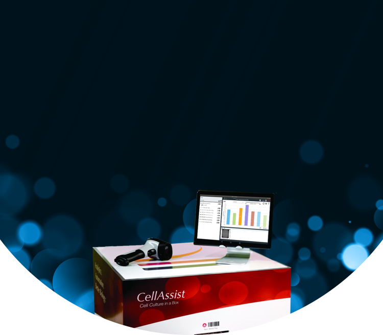
CellAssist Benchtop Imager

Single Plate Live Cell Imager

Contact Us

Bringing Cell Culture into the Data-Driven Future

An environmentally-controlled, benchtop system that images single cell culture plates from multiple users. The imaging system provides bright-field, phase contrast, and Thrive Phase Imaging at 4x, 10x, and 20x at 100+ focal planes on 6- well through 384-well flat and round bottom plates. The CellAssist Software Suite automatically builds a centralized database of images, metrics, and methods and materials.

Video: The CellAssist Experience

The "CellAssist Experience” video (3:38 minutes) provides an overview of the CellAssist Family of Systems, which combine an advanced imaging system with intuitive software to provide high-resolution images with excellent accuracy. The unique capabilities outlined in the video enable biologists to gain a deeper insight into cell dynamics, and greatly improve the reproducibility of cell, tissue, and suspension culture experiments. Learn more about the CellAssist Family of Systems and their capabilities with this video.

Transforming Cell Imaging into Rich, Reproducible Data with Ease

Enhanced Data Quality

Image single plates automatically, significantly increasing your data collection capacity and saving you valuable time that would otherwise be spent on manual tasks. Superior image registration allows for enhanced tracking and analysis of live cells frequently over time.

Remote Data Access

The CellAssist Benchtop Imager provides researchers with real-time access to cell monitoring data, allowing them to work remotely, schedule tasks, and perform analyses from anywhere. The system's 24/7 automation also eliminates the need for overtime, ensuring round-the-clock operation without additional manual effort.

Reproducibility and Traceability

Ensure consistency across experiments with precise data capture and centralized storage, making it easy to reproduce and trace every step of your workflow.

Streamlined Workflows

Minimize hands-on time with fully automated image capture, analysis, and documentation, freeing time to focus on core biological experiments and allowing for 24/7 walkaway time series automation.

User-Friendly Operation

The intuitive interface and easy benchtop integration into existing lab environments make the CellAssist Benchtop Imager a seamless addition to any research setting, ensuring a smooth user experience from setup to data analysis.

Time-Efficient Automation

By automating the entire imaging process, the CellAssist Benchtop Imager significantly reduces manual intervention. This allows researchers to conduct more experiments in less time, enhancing their efficiency and productivity. With a high degree of accuracy and confidence, researchers can focus on their core biological experiments.

CellAssist Benchtop Workflow

Step 1

Capture methods and materials

Step 2

Insert plate into CellAssist Benchtop Imager

Step 3

Capture, view, and transmit to database

Step 4

Remove plate for continued workflow

Step 5

Analyze, compare, and share Images

CellAssist Benchtop Features

Key Features

  • Barcode tracking of culture plates, reagents, and workflow
  • Imaging of adherent cells, suspension cells, and organoids in standard 6-well through 384-well flat or round bottom plates
  • Scanning modes include center-of-well, standard-well, whole-well, and regions of interest
  • Wireless, and wired network connection between CellAssist instrument(s), and EvalCore Analysis Workstation, enabling location installation flexibility
  • Central database of images, and metrics, including growth rates, confluence, colony count, and size, and monoclonality
  • Detailed viewing of stitched images at varying z-height depths along with zoom, measurement, and export capabilities
  • Time lapse viewing capability to visualize colony formation, morphology, differentiation, and monoclonality

Instrument Specifications

  • Microscopy: 4x, 10x, 20x; phase contrast, bright-field, Thrive Quantitative Imaging (proprietary and enhanced quantitative phase imaging)
  • Image Field of View: 5.3mm x 4.0mm at 4x; 2.1mm x 1.6mm at 10x; 1.1 mm x 0.8 mm at 20x
  • Resolution: 2.1 μm/pixel at 4x; 0.84 μm/pixel at 10x; 0.41 µm/pixel at 20x
  • Focal Planes: Captures images at 100+ focal planes, 2 μm to 50 μm apart (user selectable), with a z-range of 4.0 mm
  • User interfaces: Tablet in laboratory, and EvalCore Analysis Workstation in laboratory or office
  • Barcode reader (Bluetooth) for plate ID, reagents, and experiment data entry
  • Tablet user interface (Wi-Fi) in laboratory for viewing, and project planning
  • Wireless access point for private network with EvalCore Analysis Workstation
  • Internal temperature: 37°C with CO2 option
  • Local storage: 4+ TB
  • Dimensions: 0.5m x 0.5m x 0.5m (lwh) / Weight: 32 kg
  • Operating environment: temp 20 to 37°C; relative humidity 20 to 80%
  • Power requirements: 100 to 240VAC 50/60Hz; 650W max

CellAssist Software & CellAssist Workstation Specifications

  • Powerful computing workstation combined with CellAssist Benchtop Imager, and CA Control software for image analysis, and project management
  • Networked to the CellAssist Benchtop Imager with either a wired or wireless connection
  • Automatic image transfer from CellAssist instrument(s)
  • Image, and video export formats: png, mp4
  • Local storage: 18TB
  • Dimensions: 0.5m x 0.2m x 0.8m (lwh) (without monitor) / Weight: 14 kg
  • Power requirements: 100 to 240VAC, 50/60Hz; 890W max

CellAssist Benchtop Benefits

Centralized Database

  • Organizes images across time, across plates, across cell lines, and across users
  • Images and displays cells at the same location on the cell culture plate within 15 μm registration across scans
  • Rapidly generates extensive data sets of 3 GB to 90 GB of images per scan

Objective metrics and quantitative reports

  • Characterizes cell dynamics, including growth curves, confluence, and the number and area of colonies
  • Analyzes and compares cells to support decision-making to reduce variability and improve quality
  • Allows comparison and optimization of multiple methods and materials

Key Workflow Details and Status of Cells

  • Accurately records and time-stamps your cell culture process with a barcode scanner
  • Captures and archives the key workflow details and the status of cells to provide confidence that data is not lost or compromised
  • Enables comparison with previous experiments

Download Brochure

Please fill out the form below for more information about Thrive Bioscience’s technologies.

"*" indicates required fields

This field is hidden when viewing the form

CellAssist Benchtop Applications

Discover how Thrive Bioscience's advanced live-cell imaging solutions are transforming research with deeper insights, greater efficiency, and unmatched accuracy across diverse applications.

Explore Applications

Questions? Let’s Talk

Connect with one of our expert staff to discuss how Thrive Bioscience technologies can enhance your cell biology research.

Contact Us

Resources

Learn more about our technologies and their use in advancing cell biology research.

RESOURCES

Video

Video: Image Differently – A Solution for Live Cell Imaging

20240101

View More

RESOURCES

Poster

In Situ Automated Live Cell Confluence and Counting

20240101

View More

RESOURCES

Technical Note

Variability in Cell Confluency: Comparison of Human and CellAssist Assessments

20240101

View More